Patient: 45 years of age, male, BCVA 0.6.
General Medical History: empty.
Ocular Medical History: in 2003 Laser-assisted in Situ Keratomileusis, in 2014 ocular blunt trauma with flap dislocation, and epithelial cell growth into the interface, in 2014 two flap-lift retreatments with surgical flap reposition, good visual recovery.
Purpose: to visualize corneal structural changes and epithelial ingrowth in a young male patient after a blunt trauma with LASIK flap dislocation after LASIK 11 years ago.
Methods: Colour Photography Anterior Segment, Anterior segment optical coherence tomography (SL-OCT), Topography (Tomey))
Findings:
Colour Photography Anterior Segment: Focal area of epithelial ingrowth.
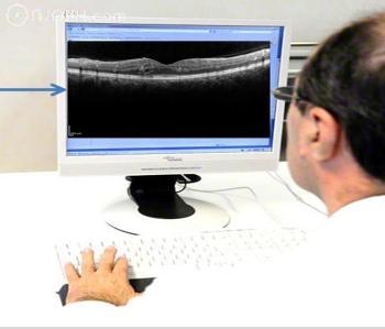
SL-OCT: epithelial ingrowth underneath the flap, no flap stromal edema.
Topography: irregular astigmatism.
Discussion:
Interface complications include infectious keratitis, diffuse lamellar keratitis, central toxic keratopathy, pressure-induced stromal keratopathy (PISK), and epithelial ingrowth. Güell J et al. (1) reported a low incidence of epithelial ingrowth after regular LASIK. From a total of 7520 LASIK refractive eyes, 13 eyes with epithelial ingrowth were treated. Randleman JB et al. (2) described epithelial ingrowth as a LASIK interface complication. Epithelial ingrowth is easily distinguishable from other interface complications and may be self-limited or require flap lift to treat irregular astigmatism and prevent flap melt.
Literature:
(1). Güell JL, Verdaguer P, Mateu-Figueras G, Elies D, Gris O, Manero F, Morral M. Epithelial ingrowth after LASIK: visual and refractive results after cleaning the interface and suturing the lenticule. Cornea. 2014 Oct;33(10):1046-50
(2) Randleman JB, Shah RD. LASIK interface complications: etiology, management, and outcomes. J Refract Surg. 2012 Aug;28(8):575-86.
-------------------------- --------------------------
-------------------------- --------------------------
-------------------------- --------------------------
-------------------------- --------------------------
-------------------------- --------------------------
-------------------------- --------------------------