| Fusiform Aneurysm of the Basilar Artery with Leftsided Trochlearis Paresis and Intermittened Diplopia (Angiography) | |
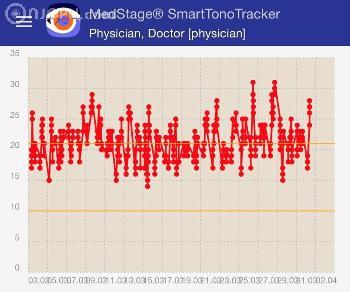
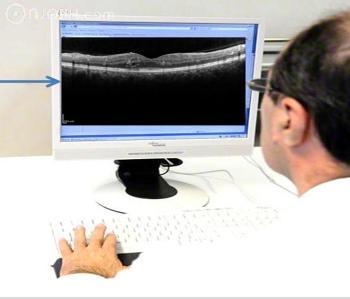
| 81 years of age, male, BCVA 0.8 at OD, 0.6 at OS Main complaint: diplopia Ocular medical history: uncomplicated cataract surgery at OD in 2009 General medical history: arterial hypertension. Purpose: to present a 81-year-old Caucasian man with a history of arterial hypertension presented to the Department of Ophthalmology complaining of diplopia. He was found to have a large basilar artery aneurysm. Methods: Ophthalmoscopy, neuro-ophthalmological examination, OCT, MRI. Findings: 1) Ophthalmoscopy showed regular retina and optic nerve head. 2) OCT showing generalized thinning of retinal nerve fiber thickness 3) Neuro-ophthalmological examination depicted left sided trochlear nerve paresis with a vertical deviation of 3.5° leading to intermittened diplopia. 4) MRI-Angiography: Imaging of the brain revealed a fusiform aneurysm of the proximal basilar artery with a diameter of 17 mm. Discussion: The visual pathways and the ocular motor cranial nerves are frequently injured by expanding cerebral aneurysms. Neuro-ophthalmologic signs and symptoms may be the only indications of an aneurysm prior to rupture. Acute or chronic visual loss may herald an aneurysm prior to rupture. Diplopia and retro-orbital pain may be warning signs that precede the discovery of a posterior communicating, basilar, or cavernous sinus aneurysm. Dolichoectasia of intracranial arteries is a rare arteriopathy characterized by elongation and widening of the arteries and disturbance of the laminar blood flow. It involves mostly vertebral and basilar arteries. In advanced cases, formation of a fusiform aneurysm is possible. Intracranial arterial dolichoectasia may be asymptomatic for a long time. However, in many cases it leads to neurological symptoms associated with haemodynamic disturbance and mass effect caused by the widened vessel.(1) Literature: (1) Baran B, Kornafel O, Guziński M, Sąsiadek M. Dolichoectasia of the circle of Willis arteries and fusiform aneurysm of basilar artery - case report and review of the literature. Pol J Radiol. 2012 Apr;77(2):54-9. | |
| Dörfler, Arnd, Prof. Dr. med., Neuroradiologie, Friedrich-Alexander-Universität Erlangen-Nürnberg, Deutschland, Erlangen, Germany; Gusek, Gabriele, Prof. Dr. med., Augenklinik, Friedrich-Alexander-Universität Erlangen-Nürnberg , Erlangen, Germany; Michelson, Georg, Prof. Dr. med., Interdisziplinäres Zentrum für augenheilkundliche Präventivmedizin und Imaging, Augenklinik, Friedrich-Alexander-Universität Erlangen-Nürnberg, Erlangen, Germany | |
| I72.5 + H49.1 | |
| Brain and Optical Pathway -> Intermittened Diplopia by Fusiform Aneurysm of the Basilar Artery (Neuro-Ophthalmological Exam, MRI-Angiography, OCT, Colour Image) | |
| Trochlear paresis, basilar aneurysma, angiography | |
| 9301 |
|
|||||||||||||||||||||||||||
|
|
Fusiform Aneurysm of the Basilar Artery with Leftsided Trochlearis Paresis and Intermittened Diplopia (Angiography)-------------------------- -------------------------- -------------------------- -------------------------- -------------------------- -------------------------- -------------------------- -------------------------- -------------------------- -------------------------- -------------------------- -------------------------- |
|
|||||||||||||||||||||||||